What’s inside a muscle?
Muscles power virtually all animal motion – so naturally there are plenty of researchers interested in understanding the connection between the form of muscles and their function. But under the surface, there is plenty of interesting hidden muscular anatomy that’s much harder to study. Many muscles are organized hierarchically, containing long, narrow cells called “muscle fibers.” Fortunately, computed tomography (CT) scans – 3-D models of anatomy resulting from images taken by a spinning X-ray beam – can allow us to visualize these individual, tiny fibers in 3-D.
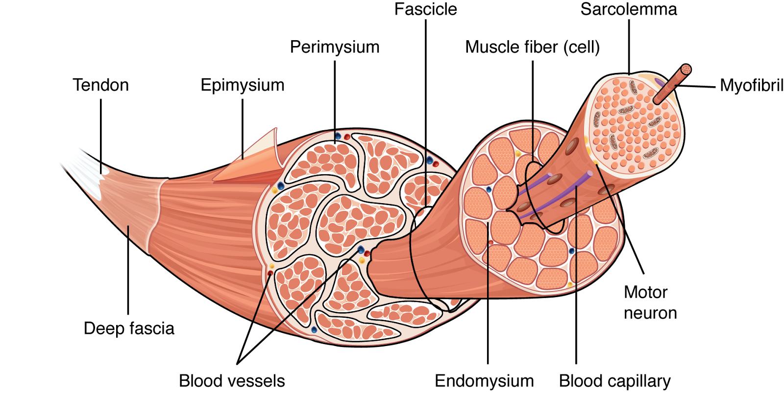
Generalized hierarchical structure of a vertebrate skeletal muscle. Source: Oregon State University
However, our only options for quantitatively analyzing muscle fibers are to (1) painstakingly trace out each one by hand, or to (2) use expensive proprietary software to separate them automatically. As a result, existing studies of internal muscle structure across the animal kingdom are few and far between – with a few notable exceptions!
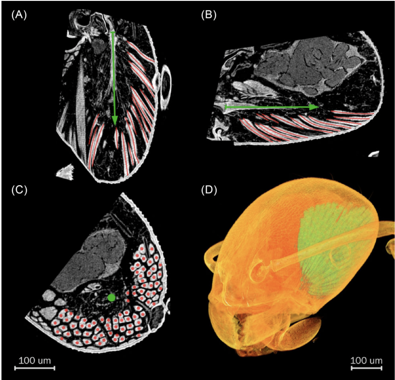
Individual muscle fibers (red) visible in a CT scan of a pharaoh ant muscle. Source: Katzke et al. (2022)
Another option…
To make this data source more accessible to a broader community of scientists, J. Katzke and colleagues from across Japan and Germany compared existing methods for tracking muscle fibers with a new, free and open-source alternative. Using each method, the researchers measured parameters like muscle area, fiber length, and number of muscle fibers from CT scans taken of both a pharaoh ant and a common starling bird. The results are published in a new article in Integrative Organismal Biology.
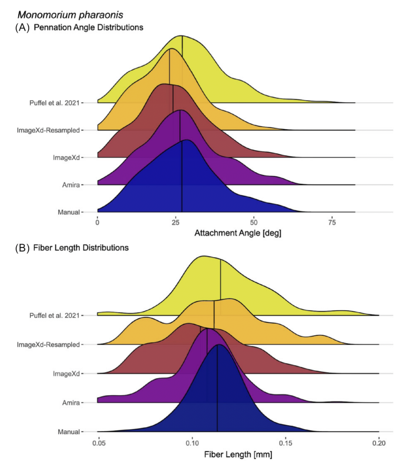
Comparison of muscle fiber analysis methods for the pharaoh ant muscle sample. Source: Katzke et al. (2022)
Despite some minor quantitative differences among the methods tested, the research team determined that all approaches are quite comparable – including the new method they developed for this study. And because this new approach relies on 3-D animation software, Katzke and colleagues were also easily able to develop beautiful visualizations of their newly acquired muscle fiber data.
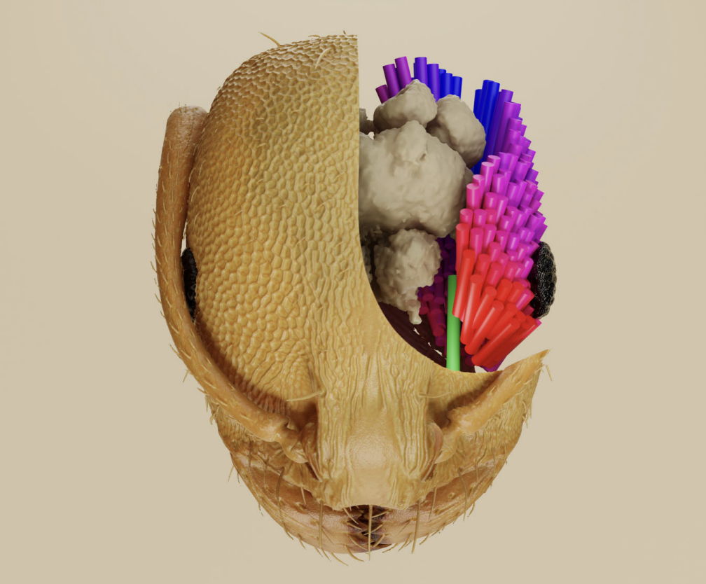
Muscle fibers in the head of a pharaoh ant, colored by orientation. Source: Katzke et al. (2022)
New opportunities
The creation of a free, automatic method for measuring muscle fiber data from CT scans makes access to this source of information a reality for many scientists across the globe. And because CT scanning, unlike dissection or other approaches, doesn’t require researchers to destroy the specimens they’re studying, this method will also make it possible to study rare species from museum collections or specimens being used for other studies.
As datasets about internal muscle structure are compiled for a broader range of animals, we will not only gain a better understanding of the diversity of muscles across the tree of life, but also of how they function differently and how they’ve evolved over hundreds of millions of years. But in the meantime, keep checking out iobopen.wordpress.com for the latest in cool, organism-centered biology!
By Armita R. Manafzadeh
Dr. Armita R. Manafzadeh studies the dynamic arthrology of the archosaur hindlimb. Her interests include functional morphology, vertebrate paleontology, and biomechanics. She is a post-doctoral fellow at Yale University starting in the fall.
