As Extracting Computed Tomography (CT) data enables virtual preservation without physically damaging the physical specimen itself, this practice is becoming increasingly common for many scientific fields. CT data can be used in many ways, such as comparing 3D structures across species, revealing the function of different structures in a body, and reconstructing movements of extinct animals. CT data can also be used in the classroom to create models used for virtual anatomical dissections or even for 3D printing specimens that would otherwise be difficult to access. The uses for CT data are vast and truly out of this world!
Today, CT data is easier to acquire due to increased access to CT scanners outside of hospitals. Many scientists have pushed to increase shared access to CT data through open-access websites such as MorphoSource and DigiMorph. Researchers and museums can scan specimens from across the universe and upload their data to these open source websites to share with others. Not only has access to CT data increased, but the development of free open-access software has allowed scientists to study that data. Horos and 3D Slicer are both examples of free open-access software that can be used to study CT data.
Despite the exciting explosion of available CT data, standardized training for CT data processing has been slower to develop. Current training protocols are usually passed along between lab members or colleagues, but a standard protocol would increase efficiency and encourage newcomers to explore the frontiers of 3D CT data. To address this problem, Buser and colleagues have provided a published workflow using only open-access software and resources.
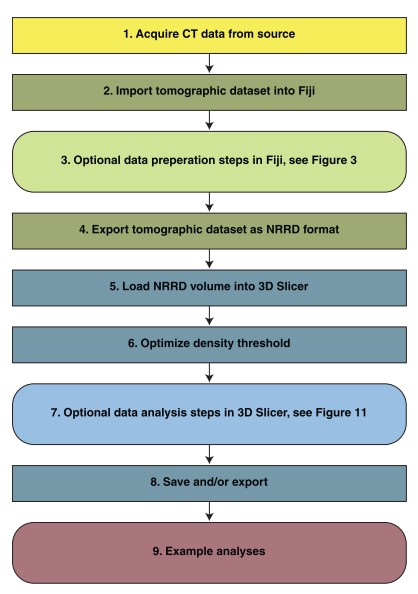
Figure 1. from Buser, et al. 2020. A flow chart listing the steps to process a CT scanned specimen.
Those interested in learning CT data processing can follow along with the 9 steps (see Figure 1, above), using step-by-step instructions. First, CT data from a specimen is downloaded, converted to an appropriate format, and imported into the open-access software, Fiji. From there, you can decide if and how you would like to prepare your data by calibrating for length measurements or isolating particular structures on the specimen. Data can then be loaded into 3D Slicer where the 3D images can be optimized and analyzed- this is where the magic happens! In 3D Slicer, you can take measurements of your specimen, add markers to do geometric morphometrics, or prepare the file for 3D printing. These methods, listed in an easy-to-follow format, can allow anyone the confidence to launch into 3D analysis.
There are so many potential benefits to using CT data for research and education. Once CT data is shared in an open-access format, it can be used for several analyses, with no time limit, and can be accessed anywhere (like in your own home!) These data can be used to expand educational materials through low-cost means such as 3D printing and virtual specimen access. Combining CT data analysis with other techniques may increase our understanding of complex physiological and anatomical systems, extinct taxa, and biomechanical attributes of the musculoskeletal system. There is still so much to learn with this technology, and with each researcher that learns these methods, we take one small step in tech, and one giant leap in open-access science!
Amanda M. Palecek-McClung is a PhD student at Clemson University. She studies functional morphology, biomechanics, and adhesion mechanisms in fish and other vertebrates. You can find more about her at ampalecek.weebly.com or contact her at apalece@g.clemson.edu.
